 3 days ago
6
3 days ago
6
ARTICLE AD BOX
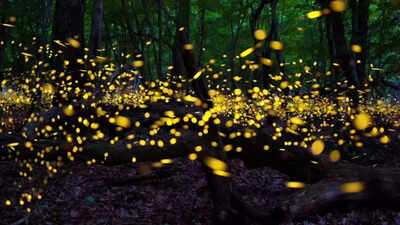
For more than a century, fireflies have been treated as biology’s most dependable natural light source. Each species was thought to emit peak light of the same intensity, an optical fingerprint determined by the structure of an enzyme called luciferase.
That assumption has shaped bioluminescence imaging (BLI), an imaging tool used to track tumours in cancer research.Researchers at Gauhati University found insects within the same species of the Indian firefly Asymmetricata circumdata do not emit identical light; their emission peaks vary measurably. The earlier theory, turns out, could have led to misreading tumour sizes and response to treatment in pre-clinical research, thereby affecting drug development too.“Positions of the emission peaks are not the same; these differ by nine nanometres,” says A J Borah, the first author of the study, “and the difference is non-negligible.” This could be because of the localised microenvironment around the firefly or slight changes in the structure of the enzyme luciferase within the species.Until now, variations in emission peaks were reported only between species and linked to evolutionary differences, reinforcing the idea that the enzyme structure remains identical within a species.
The new research also opens questions on the interpretation of biological light signals.
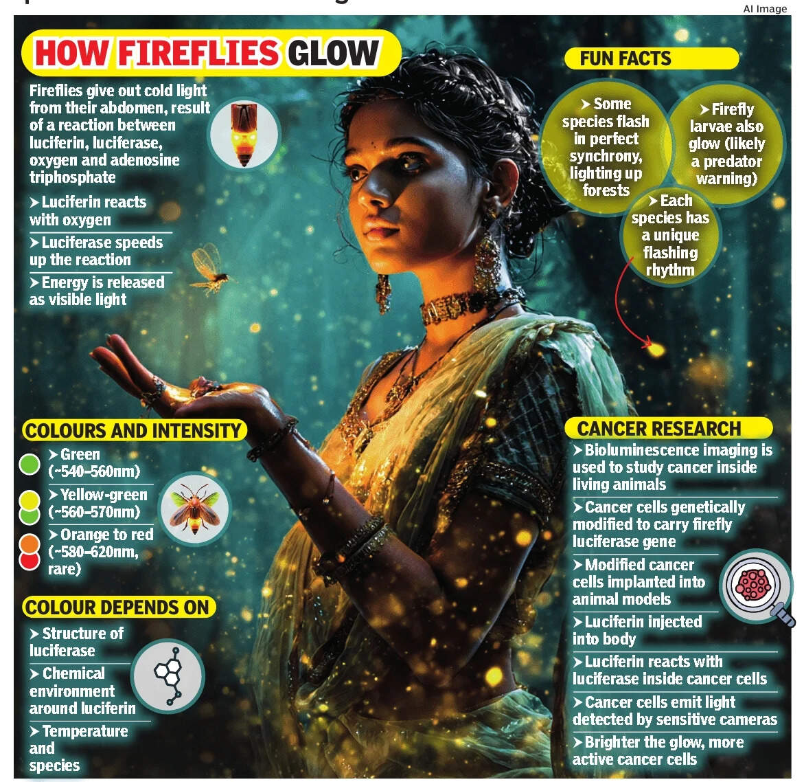
Inside the glowFirefly light is produced through a chemical reaction involving organic substance luciferin, enzyme luciferase, oxygen, adenosine triphosphate (ATP) and the cell’s energy currency. The reaction creates an excited molecule called oxyluciferin, which emits light as it returns to a lower-energy state. The colour of that light depends on how energy is arranged within the molecule.Gauhati University researchers recorded light emission from 70 male fireflies over three summers, analysing their natural flashes at normal temperatures. Instead of seeing a single, fixed peak for the species, they found emission values ranging from 561 to 570 nanometres, clustering around an average of 565 nanometres (nm).From a spectroscopy point of view, this spread shows that oxyluciferin molecules are behaving in different ways across individuals.
“As these are molecular spectra, a shift of 2–3nm can be considered negligible. But a shift of 9nm is not,” says Anurup Gohain Barua, corresponding author of the study.Luciferase effectIn bioluminescence imaging, cancer cells are genetically engineered in the laboratory to carry the gene for luciferase, the light-producing enzyme originally derived from fireflies. This is done by inserting the luciferase gene into the cancer cell’s DNA using molecular tools such as viral vectors or plasmids, so that the cell continuously produces the enzyme as it grows and divides.Once these modified cancer cells are implanted into an animal model, researchers administer luciferin, the chemical substrate for luciferase, usually by injection into the bloodstream or into the abdominal cavity. Luciferin circulates through the body and enters the cancer cells, where it reacts with luciferase in the presence of oxygen and cellular energy (adenosine triphosphate). This reaction produces visible light.The emitted light is faint, but can be detected by highly sensitive cameras, allowing scientists to track tumour growth, spread, and response to treatment in real time, without invasive procedures. The brightness of this glow is usually taken as a direct measure of the size of the tumour, and that interpretation rests on the assumption that luciferase always produces light with a stable and predictable colour.Prof Subhradip Karmakar of the department of biochemistry at AIIMS Delhi says the firefly study highlights a gap in that thinking.
“Different wavelengths travel through tissues differently,” he explains. Red and nearinfrared light can pass deeper through tissue, while green light is absorbed more easily. “A weak signal might not mean fewer cancer cells. It could simply be a wavelength that does not pass well from that tumour’s location.
”This means changes in emission colour caused by enzyme structure or the local cellular environment could affect how the tumour size or drug response is judged.
“The same number of cancer cells could appear brighter or darker depending on where they are and what wavelength they emit,” says Karmakar.The issue becomes more complicated when cancer spreads to multiple organs. Apparent differences in treatment response between tumour sites may not always reflect real biological changes. “We might think that one metastasis is responding better to treatment when it’s really just sending out a wavelength that goes deeper into the tissue,” he says.Opportunity in challengeThe Gauhati University authors are careful not to overstate their findings. “This is not conclusive, it’s only a suggestion,” says Barua. For imaging science, this points to the need for better calibration.Karmakar compares current practice to using a detector tuned to just one colour. “If you’re using a light meter that can see only red light, and if the light turns orange, the readings will be wrong. That’s pretty much what’s going on here,” he says.
Future imaging systems, he suggests, may need to detect a wider range of light or adjust measurements based on the tumour environment.At the same time, this sensitivity could be turned into an advantage. Small wavelength shifts could help create ‘smart’ probes that respond to specific conditions inside tumours or to drug delivery.“This involves creating luciferase variants that are sensitive enough to pick up what we want, like tumour growth or drug effectiveness, but stable enough that they don’t give false signals from normal biological changes,” says Karmakar.Globally, bioluminescence imaging remains a powerful tool in cancer research because of its high sensitivity. It can detect very small numbers of cancer cells long before conventional imaging methods such as MRI or CT scans, and without radiation exposure. The firefly study does not change this overnight. But by revealing unexpected variation in one of biology’s most trusted light sources, it prompts scientists to ask how much more is happening inside this glow than they once believed.


 English (US) ·