

.png)
.png)
.png)
.png)


ARTICLE AD BOX
Last Updated:July 28, 2025, 17:49 IST
UP Woman’s Foetus Growing In Liver, Not Uterus: Only 8 such cases reported worldwide. Meerut doctors say this could be India’s first intrahepatic ectopic pregnancy
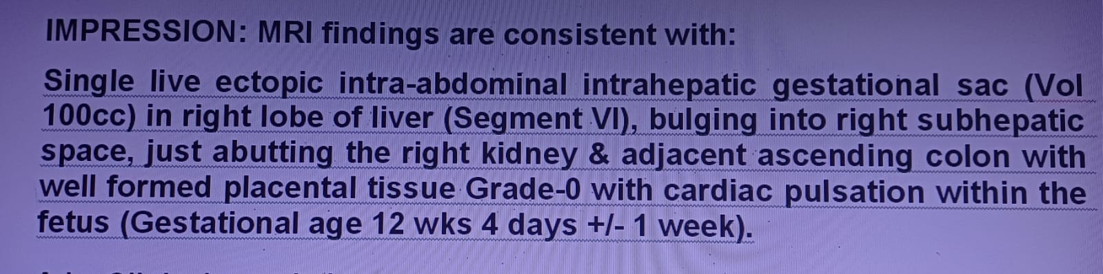
The MRI scans. (News18)
Doctors in Uttar Pradesh’s Bulandshahr were in for a shock when an MRI scan of a 30-year-old woman revealed that she was 12 weeks pregnant, but in her liver, instead of uterus.
The rare medical condition, known as intrahepatic ectopic pregnancy, has left not only the couple but the medical community astonished, with experts suggesting this could be the first such case ever reported in India.
“When I saw the scan, I could not believe my eyes. The foetus was embedded in the right lobe of the liver, and there were clear cardiac pulsations. I have never seen such a case in my career, and according to available data, this might be India’s first intrahepatic ectopic pregnancy," said Dr KK Gupta, a radiologist at a private imaging centre in Meerut, who uncovered it while carrying out the MRI abdomen test of the woman.
How was the diagnosis done?
The turning point in the case came when the woman, after weeks of abdominal pain and vomiting, was referred for an MRI of the abdomen — a test often used when ultrasound or routine scans fail to explain symptoms.
The MRI was performed at a private imaging centre in Meerut under the supervision of Dr KK Gupta, a senior radiologist with decades of experience in advanced imaging. Unlike a routine ultrasound, the MRI provided high-resolution, layered images of the abdominal organs.
According to Dr Gupta, the scan revealed a startling anomaly. “We observed a well-formed gestational sac inside the right lobe of the liver. The foetus measured approximately 12 weeks in gestational age. Most strikingly, the scan confirmed active cardiac pulsations, establishing that the foetus was alive. At the same time, the uterus was completely empty, ruling out a normal intrauterine pregnancy," Dr Gupta explained.
He further detailed that the foetus appeared embedded deep into the parenchymal tissue of the liver, with blood vessels from the organ supplying nutrition to the sac. This confirmed that the pregnancy had implanted directly into the hepatic tissue — an occurrence almost unheard of in India.
Dr Gupta also said that the diagnosis was double-checked by repeating certain MRI sequences to rule out imaging errors. “Initially, I even thought it might be an imaging artifact. But repeated scans, taken from different planes, confirmed the presence of a live foetus within the liver tissue itself. At that moment, we realised we were dealing with an extremely rare, high-risk pregnancy," he added.
Only 8 cases of liver pregnancies reported so far
Pregnancies outside the uterus, or ectopic pregnancies, are uncommon, accounting for 1–2% of all pregnancies. Most — about 97% — occur in the fallopian tubes. Rare cases are found in the ovaries or the abdominal cavity. But intrahepatic implantation — when the fertilised egg attaches to the liver — is one of the rarest forms known in medical science.
According to published literature, only eight cases of intrahepatic ectopic pregnancy have been reported worldwide so far, in countries including China, Nigeria, the United States, and parts of Europe.
Why is it dangerous?
The liver is one of the most vascular organs in the body, with an extensive blood supply. While this allows the foetus to receive nourishment temporarily, it also poses an enormous risk to the mother. The growing foetus can cause liver rupture or massive hemorrhage if not treated promptly.
Dr Jyotsna Mehta, a renowned gynaecologist and obstetrician from Lucknow, explained why this case is particularly alarming: “This is a once-in-a-lifetime case for most doctors. The liver’s rich blood supply can sustain foetal growth initially, but it puts the mother in grave danger. Removing the foetus is extremely risky — even a minor surgical slip can lead to uncontrolled haemorrhage. The immediate priority is the mother’s survival. The pregnancy, unfortunately, cannot continue safely."
She added that in similar cases worldwide, doctors sometimes attempt to remove the foetus surgically while leaving the placenta attached, later shrinking it with medication to reduce blood loss. “Each decision has to be highly individualised, and such surgeries demand extraordinary coordination between radiologists, gynaecologists, and liver surgeons," she said.
India’s first documented case?
The Meerut case could be India’s first reported instance of intrahepatic ectopic pregnancy. “Based on international literature, there is no record of such a pregnancy being reported from India before. Documenting this case will be vital, as it can help the global medical community understand, prepare for, and manage similar rare conditions in the future," Dr Gupta added.
The patient’s current condition
The woman is currently under strict medical supervision while doctors chart the safest course of treatment. A multidisciplinary team, including obstetricians, hepatobiliary surgeons, radiologists, and anaesthesiologists, has been assembled to plan a complex surgery.
While the fate of the patient remains uncertain until surgery is successfully performed, one fact is already clear: this rare diagnosis has carved its place in Indian medical history as a case that will be studied and remembered for years to come.
FAQs on intrahepatic ectopic pregnancy
What is it?
A rare form of ectopic pregnancy where the fertilised egg implants and grows inside the liver instead of the uterus.
How rare is it?
Extremely rare — only eight cases documented worldwide so far. The Meerut case may be India’s first.
Why is it dangerous?
The liver has an extensive blood supply. Any rupture or surgical attempt risks massive internal bleeding, which can be life-threatening.
What are the symptoms?
- Persistent abdominal pain
- Nausea and vomiting
- Weakness or dizziness
- Abnormal bleeding
- No relief from routine treatment
How is it diagnosed?
Through high-resolution imaging, especially MRI scans, since routine ultrasounds may miss the anomaly.
What are the treatment options?
Emergency surgery to remove the foetus (often requiring part of the liver to be removed)
Placenta sometimes left attached and shrunk with medication to control bleeding
Priority always remains saving the mother’s life.
view comments- Location :
- First Published:
News india UP Woman’s Foetus Growing In Liver, Not Uterus: What Is Rare Intrahepatic Ectopic Pregnancy?
Disclaimer: Comments reflect users’ views, not News18’s. Please keep discussions respectful and constructive. Abusive, defamatory, or illegal comments will be removed. News18 may disable any comment at its discretion. By posting, you agree to our Terms of Use and Privacy Policy.


 English (US) ·